
Abdominal Separation in the Pre and Postnatal period
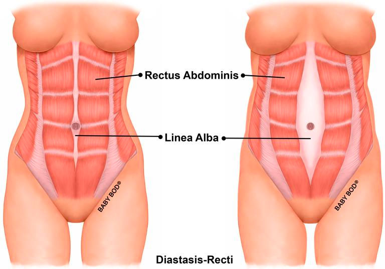
Abdominal separation (medically termed Diastasis Rectus Abdominis) is the very common separation of the abdominal muscles during the term of a pregnancy. Principally, it is the separation of the rectus abdominis muscle which runs in two parallel bands down the centre of the stomach. This muscle attaches from the bottom of the breastbone to the pubic bone.
As the womb expands during pregnancy the rectus abdominis parts down the middle to allow more space for the growing baby.
How does abdominal separation occur?
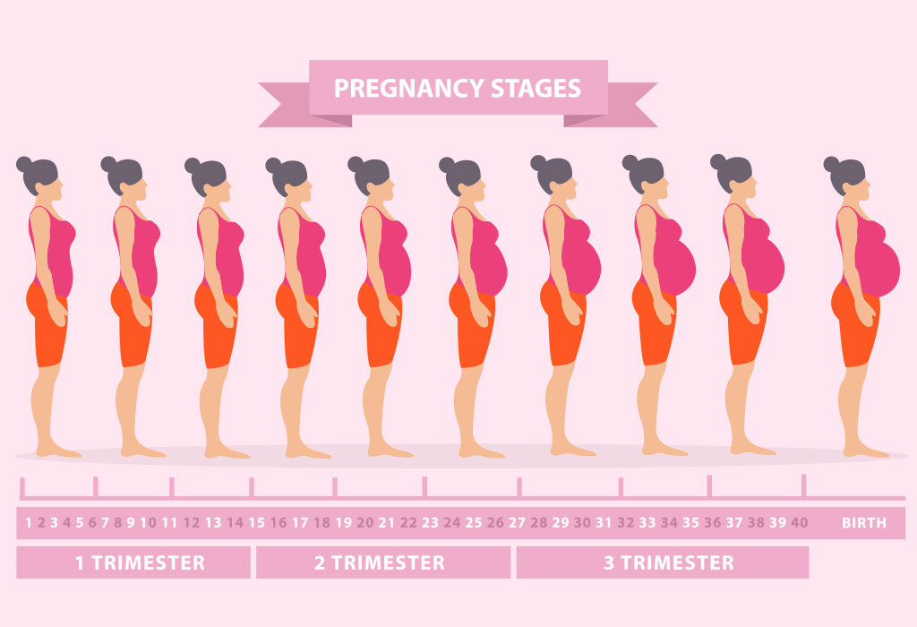
As a woman progresses through her pregnancy, there are two factors that allow for the separation of the abdominal muscles, i.e. Diastasis Rectus Abdominis (DRA), to occur:
1. Hormonal changes:
The natural release of hormones, such as relaxin, oestrogen and progesterone, causes the softening of the fibrous connective tissue that attaches the abdominal muscles down the midline, called the linea alba.
2. Growing womb:
As the baby grows bigger and heavier, the growing womb pushes out and down against the abdominal muscles. This increased load then causes the already softened connective tissue to gradually stretch, and the abdominal muscles to creep apart.
How common is abdominal separation?
In research, the prevalence of abdominal separation in pregnancy varies widely, anywhere between 30-100% of women. This is because there are a variety of methods used to measure the distance between the abdominal muscles. However, we do know that abdominal separation or DRA is very common, even up to 1year after birth.
One study found that(1):
- At 6 weeks after birth, 60% of women had abdominal separation over 2 finger-widths
- At 6 months after birth – 45% of women
- At 1 year after birth – 33% of women
How is it diagnosed?
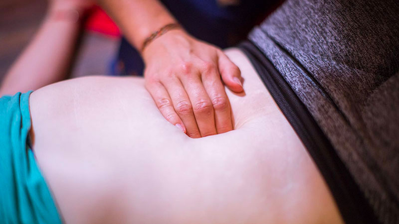
Abdominal Separation can be checked by a GP, mid-wife or physiotherapist.
The degree of abdominal separation, or distance between the rectus abdominis muscle bands, is measured clinically by finger-widths, tape measure or ultrasound(2). Commonly, measurements will be taken along the length of the muscle, both at rest and during an abdominal contraction.
Abdominal Separation and Low Back Pain
Historically, abdominal separation or DRA was feared as it was thought to increase risk of low back pain, urinary incontinence and pelvic organ prolapse. However, studies have found no correlation between low back pain and incontinence with abdominal separation. There may be a small association with abdominal separation and prolapse(3).

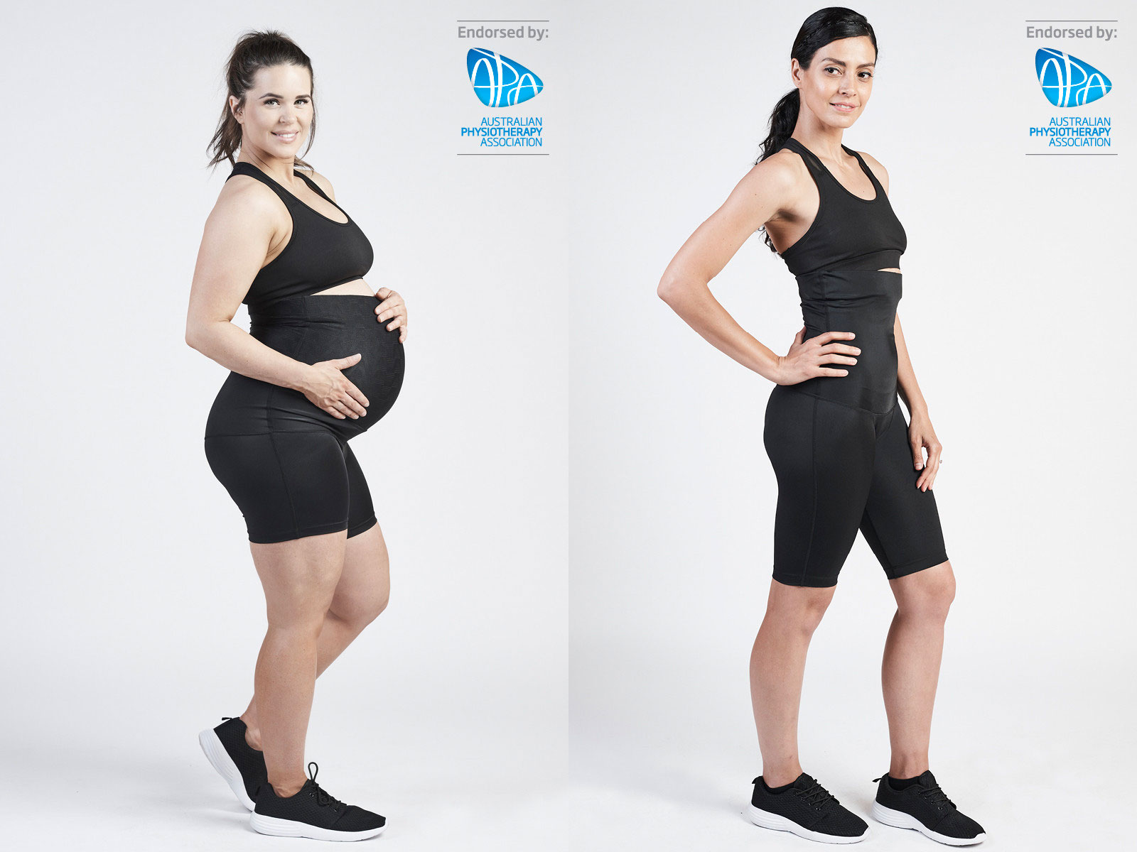
Treatment of Abdominal Separation
Woman are recommended to visit a Women’s Health Physiotherapist if they are encountering issues related to core strength and stability, or wish to improve the separation of the abdominal muscles. Early on after pregnancy, particularly in the first 6 months, is the key time to improve abdominal separation.
During pregnancy, abdominal separation appears to be quite normal. One study finding that women at 35weeks pregnant had an average separation of 6cm4! However, if abdominal separation is significant or problematic during pregnancy, supportive garments may be worn during the pregnancy to minimise the separation.
Depending on a woman’s goals and level of separation, treatment may involve:
- An individualised abdominal strengthening program prescribed by a Physiotherapist
- Supporting garments, such as abdominal binders, shapewear and compression garments
- Surgical repair
References
1. Sperstad JB, Tennfjord MK, Hilde G, Ellström-Engh M, Bø K. Diastasis recti abdominis during pregnancy and 12 months after childbirth: prevalence, risk factors and report of lumbopelvic pain. Br J Sports Med. 2016 Sep;50(17):1092-6. doi: 10.1136/bjsports-2016-096065. Epub 2016 Jun 20. PubMed PMID: 27324871; PubMed Central PMCID: PMC5013086.
2. Keeler, Jessica & Albrecht, Melissa & Eberhardt, Lauren & Horn, Laura & Donnelly, Chantal & Lowe, Deborah. (2012). Diastasis Recti Abdominis. Journal of Womenʼs Health Physical Therapy. 36. 131-142. 10.1097/JWH.0b013e318276f35f.
3. Benjamin DR, Frawley HC, Shields N, van de Water ATM, Taylor NF. Relationship between diastasis of the rectus abdominis muscle (DRAM) and musculoskeletal dysfunctions, pain and quality of life: a systematic review. Physiotherapy. 2019 Mar;105(1):24-34. doi: 10.1016/j.physio.2018.07.002. Epub 2018 Jul 24. PubMed PMID: 30217494.
4. Mota P, Pascoal AG, Carita AI, Bø K. Normal width of the inter-recti distance in pregnant and postpartum primiparous women. Musculoskelet Sci Pract. 2018 Jun;35:34-37. doi: 10.1016/j.msksp.2018.02.004. Epub 2018 Feb 20. PubMed PMID: 29494833.